Mean Pre-op corneal astigmatism
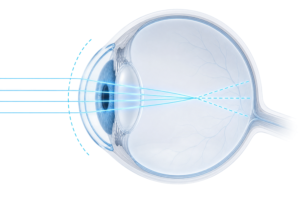 0.78D
0.78D


A real-world single-surgeon case series of eyes with mild corneal astigmatism (mean 0.78D) implanted with the 1.25D enVista Aspire Toric
Show Case Details
Mild corneal astigmatism sits in a real decision gap. Below roughly 1D the cylinder is often left uncorrected, yet residual refractive cylinder as low as 0.5D can measurably compromise uncorrected vision.
The practical question is whether the lowest-power toric option meaningfully reduces astigmatism and minimizes residual cylinder. Here is what one real-world case series found.
In an early case series of eyes with mild corneal astigmatism, the lowest-power enVista Aspire Toric (1.25D) reduced residual refractive cylinder to approximately 0.35D, with 20/25 or better uncorrected distance vision in 89%.
Early case series — 9 eyes (8 patients), single surgeon, retrospective, short follow-up.
Stephenson case series · All eyes implanted with the 1.25D enVista Aspire Toric
Two distinct measures, shown side by side.
0.78D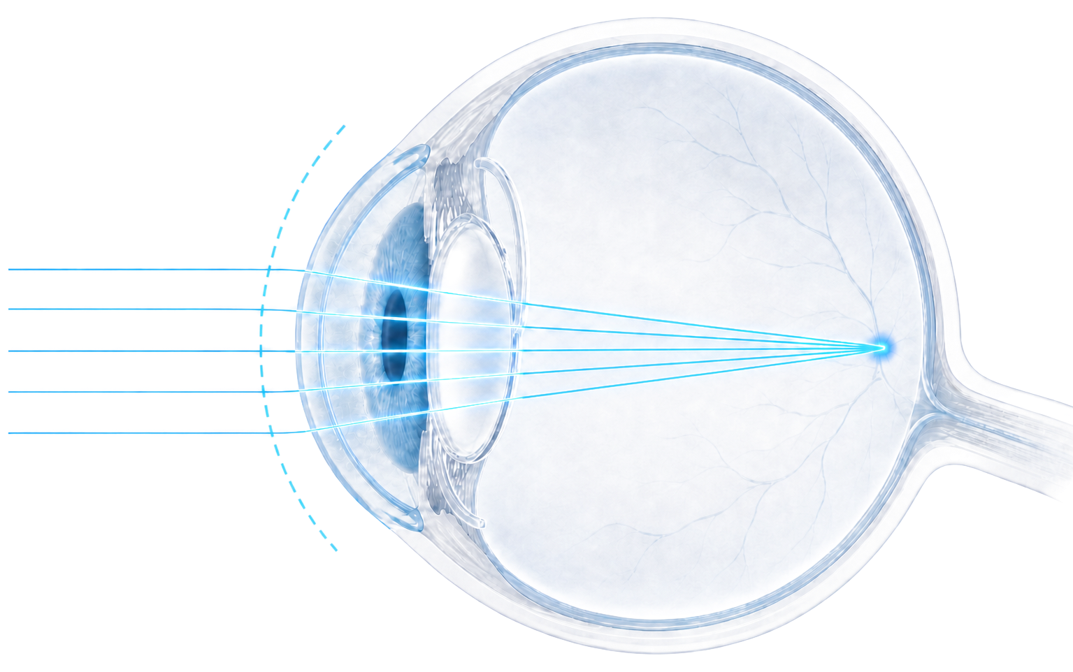 0.35D
0.35DSource: Stephenson, ASCRS 2025, pp. 6 & 8
The achieved refraction landed on the intended emmetropic (plano) target.
Source: Stephenson, ASCRS 2025, pp. 5, 6 & 8
Dominant percentages surface the key clinical success rates.

20/20 or better

20/25 or better

20/32 or better
Source: Stephenson, ASCRS 2025, p. 7
See the full Stephenson presentation, including the residual-cylinder and visual-acuity data above.
 Download Case Series PDF
Indications
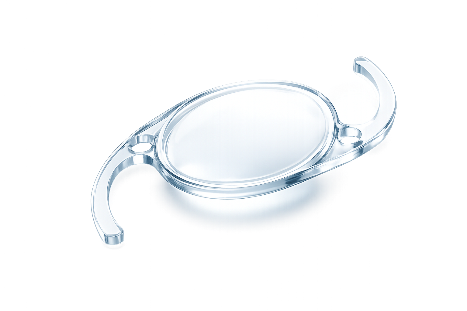
The enVista Aspire™ toric hydrophobic acrylic IOL (non-preloaded model ETA) is indicated for primary implantation in the capsular bag of the eye in adult patients for the visual correction of aphakia and corneal astigmatism following the removal of a cataractous lens for improved uncorrected distance vision.
Device Description
The Aspire IOL uses an optical modification of the posterior aspheric surface to create a small continuous increase in IOL power within the central 1.5 mm diameter to slightly extend the depth of focus. However, clinically meaningful extension of the depth of focus has not been demonstrated in clinical trials.
Warnings
Physicians considering IOL implantation under any of the following circumstances should weigh the potential risk/benefit ratio: (1) Recurrent severe anterior or posterior segment inflammation or uveitis; (2) Patients in whom the IOL may affect the ability to observe, diagnose, or treat posterior segment diseases; (3) Surgical difficulties at the time of cataract extraction, which might increase the potential for complications (e.g., persistent bleeding, significant iris damage, uncontrolled positive pressure, or significant vitreous prolapse or loss); (4) A distorted eye due to previous trauma or developmental defect in which appropriate support of the IOL is not possible; (5) Circumstances that would result in damage to the endothelium during implantation; (6) Suspected microbial infection; (7) Patients in whom neither the posterior capsule nor zonules are intact enough to provide support; (8) Rotation of the IOL away from the intended axis can reduce its astigmatic correction. Misalignment greater than 30° may increase postoperative refractive cylinder. If necessary, IOL positioning should occur prior to capsule fibrosis and IOL encapsulation.
Precautions
Neither the safety and effectiveness, nor the effects of the Aspire IOL optical design on depth of focus, contrast sensitivity, and subjective visual disturbances (glare, halo, etc.) have been evaluated clinically. MTF testing of the Aspire IOL optical design (used in model ETA) may aid the surgeon in understanding the theoretical image quality expected with the Aspire IOL compared to the enVista monofocal IOL MX60E. However, these do not fully assess all aspects of clinical difficulties under all conditions. Surgeons must weigh the potential benefits of the modified optical design of the Aspire IOL (model ETA) against the potential for risks associated with a degradation in vision quality and the lack of clinical data to characterize the impact of the Aspire IOL optical design on contrast sensitivity and subjective visual disturbance. These considerations may be especially relevant to patients with certain pre-existing ocular conditions (prior corneal refractive surgery, irregular corneal astigmatism, severe corneal dystrophy, macular disease, optic nerve atrophy, etc.) or intraoperative conditions (posterior capsular rupture, complications in which the IOL stability could be compromised, inability to place IOL in capsular bag, etc.).
The safety and effectiveness of the IOL have not been substantiated in patients with pre-existing ocular conditions and intraoperative complications. Careful preoperative evaluation and sound clinical judgment should be used by the surgeon to decide the benefit/risk ratio before implanting an IOL in a patient with one or more of these conditions. Physicians considering IOL implantation in such patients should explore the use of alternative methods of aphakic correction and consider IOL implantation only if alternatives are deemed unsatisfactory in meeting the needs of the patient.
Patients with preoperative problems, such as corneal endothelial disease, abnormal cornea, macular degeneration, retinal degeneration, glaucoma, and chronic drug miosis may not achieve the visual acuity of patients without such problems. The physician must determine the benefits to be derived from IOL implantation when such conditions exist.
Adverse Events
As with any surgical procedure, there is risk involved. Potential complications accompanying cataract or implant surgery may include, but are not limited to the following: corneal endothelial damage, infection (endophthalmitis), retinal detachment, vitritis, cystoid macular edema, corneal edema, pupillary block, cyclitic membrane, iris prolapse, hypopyon transient or persistent glaucoma, and secondary surgical intervention. Secondary surgical interventions include but are not limited to: lens repositioning, lens replacement, vitreous aspiration or iridectomy for pupillary block, wound leak repair, and retinal detachment repair.
CAUTION: Federal law restricts this device to sale by or on the order of a physician.
ATTENTION: This is not all you need to know. Please refer to the Directions For Use labeling for a complete listing of indications, full risk and safety information, clinical study information, etc.
Stephenson D. Retrospective Case Series of Patients Implanted with a Low-Powered Toric Monofocal IOL with an Enhanced Optic. ASCRS 2025, Presented 110177.
Dee Stephenson, MD, reported research, consulting, advisory and speaking relationships, including Bausch + Lomb and Zeiss. Medical writing support was provided by Panacea 85 Ltd, funded by Bausch + Lomb.